欢迎来到国际知识中心
Scientists create miniature human heart model in the lab

Cardiovascular-related disorders are a significant global health problem. Cardiovascular disease (CVD) is the leading cause of death in developed countries, accounting for a third of the mortality rate in the United States alone. At the same time, congenital heart defects (CHD) affect less than 1 percent of newborns, making it the most common congenital disabilities in humans.
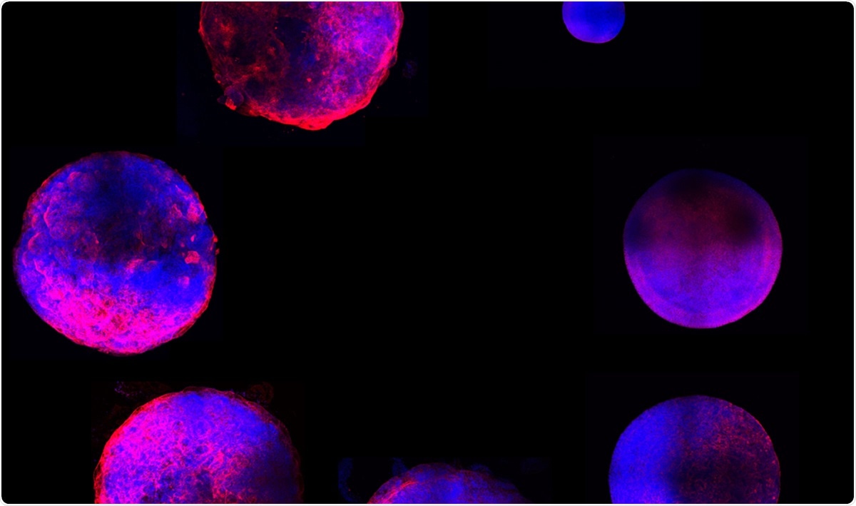
Studying cardiovascular disorders is a common problem among scientists, but now, a team of researchers from Michigan State University has created a miniature human heart model in the laboratory. For the first time in history, this mode is complete with all primary heart cell types and a functioning structure of chambers and vascular tissue.

The scientists believe that their model can enable them to study all kinds of cardiac disorders more accurately than before.

Appearing on the preprint server bioRxiv*, the study was funded by grants from the American Heart Association and the National Institutes of Health.

Human heart organoids (hHO)

Research on stem cells and developmental biology has made it possible to grow small pieces of tissue in the laboratory called organoids. Organoids closely resemble many organs, from the liver and brain to the kidneys.

Getting human tissue to study is difficult to obtain, but organoids provide scientists with new alternatives to study human organs and disease. They can use organoids to study the complex interactions of cells, which is not possible with other experimental models.

Human heart organoids (hHOs) are mini hearts that constitute powerful models to study all kinds of cardiac disorders. The hHOs were created by a novel stem cell framework that is based on the embryonic and fetal developmental environments.

These organoids are self-assembling 3D constructs that mimic organ properties and structures, allowing scientists to study heart diseases more efficiently and more accurately at the cellular level.

Real-time analysis and observation

Further, since the organoids developed following natural cardiac embryonic development, the researchers were able to study the natural growth of an actual fetal human heart in real-time. This allows them to learn more about the heart’s structure and function.

One of the hurdles experienced by scientists in studying congenital heart defects is access to a developing heart. In the past, they study fetal heart development with the use of animal models or donated fetal remains. Now, with a mini heart that they can observe from the start of its development, it can provide more accurate information on how congenital heart defects emerge.

Future plans

For the research team, the study is just the first of the many experiments they will make. While their model is complex, it is far from perfect. The team plans on improving the organoid to provide a better version for future research and use.

The team said that the mini heart is not as perfect as the human heart, but they are working on it. Also, the team hopes for a broad range of applicability of the miniature hearts, enabling them to study other cardiovascular-related diseases, including chemotherapy-induced cardiotoxicity and the effects of diabetes on the developing fetal heart during pregnancy.

*Important Notice

bioRxiv publishes preliminary scientific reports that are not peer-reviewed and, therefore, should not be regarded as conclusive, guide clinical practice/health-related behavior, or treated as established information.

Original Text (This is the original text for your reference.)

Cardiovascular-related disorders are a significant global health problem. Cardiovascular disease (CVD) is the leading cause of death in developed countries, accounting for a third of the mortality rate in the United States alone. At the same time, congenital heart defects (CHD) affect less than 1 percent of newborns, making it the most common congenital disabilities in humans.

Studying cardiovascular disorders is a common problem among scientists, but now, a team of researchers from Michigan State University has created a miniature human heart model in the laboratory. For the first time in history, this mode is complete with all primary heart cell types and a functioning structure of chambers and vascular tissue.

The scientists believe that their model can enable them to study all kinds of cardiac disorders more accurately than before.

Appearing on the preprint server bioRxiv*, the study was funded by grants from the American Heart Association and the National Institutes of Health.

Human heart organoids (hHO)

Research on stem cells and developmental biology has made it possible to grow small pieces of tissue in the laboratory called organoids. Organoids closely resemble many organs, from the liver and brain to the kidneys.

Getting human tissue to study is difficult to obtain, but organoids provide scientists with new alternatives to study human organs and disease. They can use organoids to study the complex interactions of cells, which is not possible with other experimental models.

Human heart organoids (hHOs) are mini hearts that constitute powerful models to study all kinds of cardiac disorders. The hHOs were created by a novel stem cell framework that is based on the embryonic and fetal developmental environments.

These organoids are self-assembling 3D constructs that mimic organ properties and structures, allowing scientists to study heart diseases more efficiently and more accurately at the cellular level.

Real-time analysis and observation

Further, since the organoids developed following natural cardiac embryonic development, the researchers were able to study the natural growth of an actual fetal human heart in real-time. This allows them to learn more about the heart’s structure and function.

One of the hurdles experienced by scientists in studying congenital heart defects is access to a developing heart. In the past, they study fetal heart development with the use of animal models or donated fetal remains. Now, with a mini heart that they can observe from the start of its development, it can provide more accurate information on how congenital heart defects emerge.

Future plans

For the research team, the study is just the first of the many experiments they will make. While their model is complex, it is far from perfect. The team plans on improving the organoid to provide a better version for future research and use.

The team said that the mini heart is not as perfect as the human heart, but they are working on it. Also, the team hopes for a broad range of applicability of the miniature hearts, enabling them to study other cardiovascular-related diseases, including chemotherapy-induced cardiotoxicity and the effects of diabetes on the developing fetal heart during pregnancy.

*Important Notice

bioRxiv publishes preliminary scientific reports that are not peer-reviewed and, therefore, should not be regarded as conclusive, guide clinical practice/health-related behavior, or treated as established information.

Comments

    Something to say?

    Log in or Sign up for free

    Disclaimer: The translated content is provided by third-party translation service providers, and IKCEST shall not assume any responsibility for the accuracy and legality of the content.
    Translate engine
    Article's language
    English
    中文
    Pусск
    Français
    Español
    العربية
    Português
    Kikongo
    Dutch
    kiswahili
    هَوُسَ
    IsiZulu
    Action
    相关新闻

    Report

    Select your report category*


    Reason*


    By pressing send, your feedback will be used to improve IKCEST. Your privacy will be protected.

    Submit
    Cancel