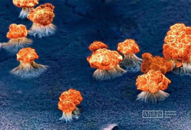
Image: Fanny Beron, École Polytechnique de Montréal, Montréal, Canada
In 2008, Fanny Beron from the École Polytechnique de Montréal and colleagues devised CoFeB nanowires. They eventually found the best composition to be Co94Fe5B1, but not before going through a long process of trial and error. While testing the magnetic properties of the films, some inevitably blew off. During one such moment, Beron used an electron scanning micrograph to record the explosion that happened when a CoFeB magnetic array was overloaded.
Subscribe to our newsletter and receive our new book for FREE
Join 50,000+ subscribers vaccinated against pseudoscience
By subscribing you agree to our
Privacy Policy. Give it a try, you can unsubscribe anytime.
Of course, there aren’t ‘mushroom clouds’, but rather solid structures that are the resting state of the array post explosion. Scanning electron microscope images are black and white, and color is added later (‘color enhanced’). This is standard practice, and the color selection thus becomes a kind of artistic expression instead of an attempt at duplicity. Still amazing!
This image won first prize in the “Science As Art” 2008 competition.
Original Text (This is the original text for your reference.)
Image: Fanny Beron, École Polytechnique de Montréal, Montréal, Canada
In 2008, Fanny Beron from the École Polytechnique de Montréal and colleagues devised CoFeB nanowires. They eventually found the best composition to be Co94Fe5B1, but not before going through a long process of trial and error. While testing the magnetic properties of the films, some inevitably blew off. During one such moment, Beron used an electron scanning micrograph to record the explosion that happened when a CoFeB magnetic array was overloaded.
Subscribe to our newsletter and receive our new book for FREE
Join 50,000+ subscribers vaccinated against pseudoscience
By subscribing you agree to our
Privacy Policy. Give it a try, you can unsubscribe anytime.
Of course, there aren’t ‘mushroom clouds’, but rather solid structures that are the resting state of the array post explosion. Scanning electron microscope images are black and white, and color is added later (‘color enhanced’). This is standard practice, and the color selection thus becomes a kind of artistic expression instead of an attempt at duplicity. Still amazing!
This image won first prize in the “Science As Art” 2008 competition.
Disclaimer: The translated content is provided by third-party translation service providers, and IKCEST shall not assume any responsibility for the accuracy and legality of the content.


 User Center
 My Training Class
 Feedback


Comments
Something to say?
Log in or Sign up for free